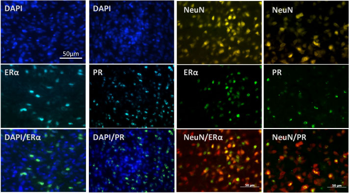
Micrographs of cells containing estrogen receptor alpha (ERα) or progesterone receptor (PR) in the medial preoptic area (mPOA) of the hypothalamus. The two columns to the left depict all cells in the mPOA stained with nucleic acid staining DAPI (in blue) and gonadal hormone receptors ERα or PR (in cyan). Qualitative analyses revealed that all staining for gonadal hormone receptors are in cells (overlay, pseudo-colored bright green), but not all cells contain steroid hormone receptors. The two columns to the right depict all neurons in the mPOA (stained with neuronal specific nuclear protein NeuN) and gonadal hormone receptors ERα or PR (in green). Qualitative analyses revealed that most hormone receptors are in neurons (overlay, pseudo-colored yellow), but not all neurons contain gonadal hormone receptors.