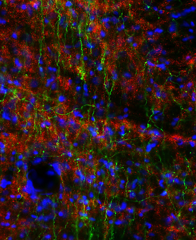
Low magnification picture taken of VTA (ventral tegmental area) fibers coming from the mPOA (green), which contained VGAT (vesicular GABA transporter; red), surrounding cells are indicated by DAPI nuclear staining (blue). Co-localization is readily visible at higher magnification (not shown here).